Medium Cardiovascular System Questions Practice Questions
Concept Explanation
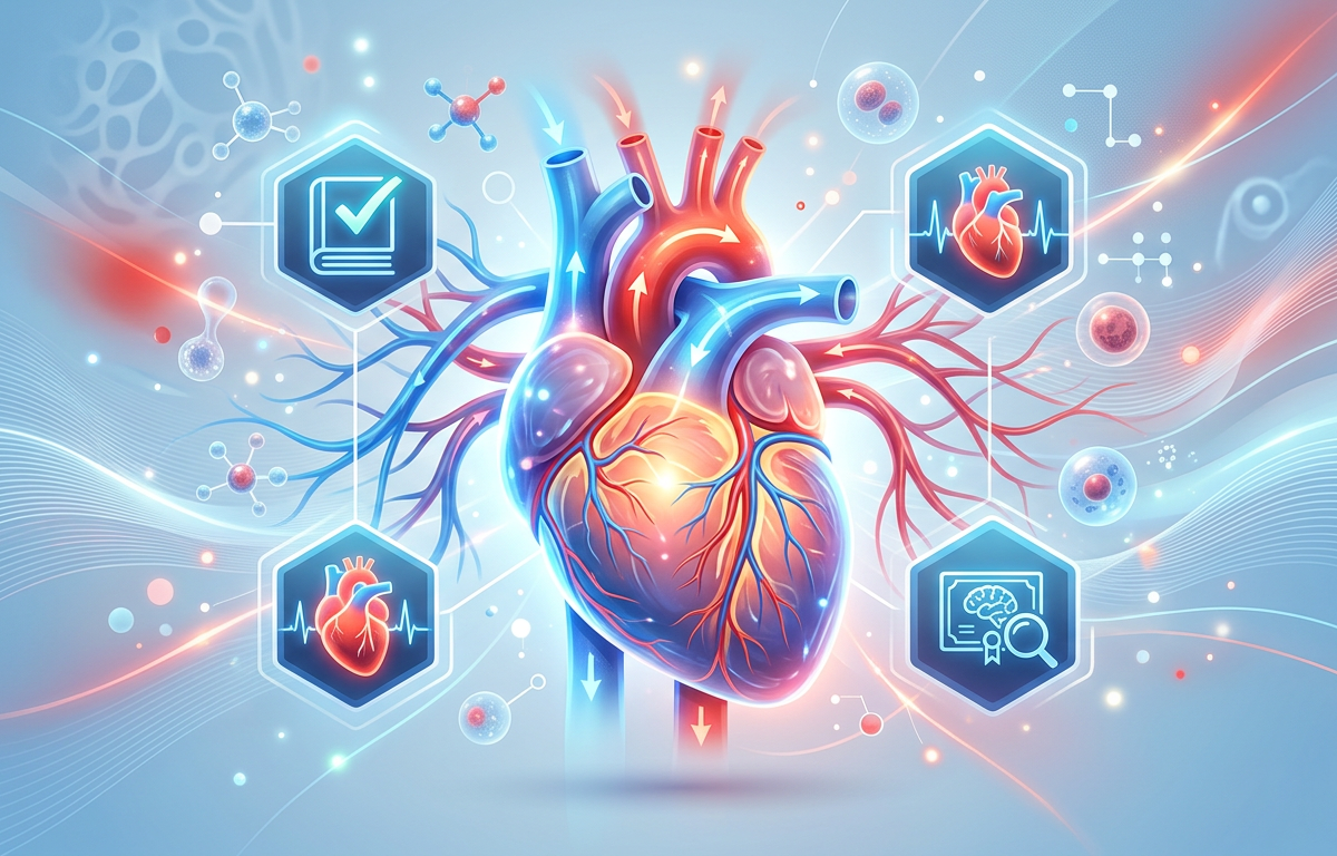
The cardiovascular system is a complex network of the heart, blood vessels, and blood responsible for transporting oxygen, nutrients, hormones, and cellular waste products throughout the body. At a medium level of understanding, focusing on Medium Cardiovascular System Questions requires a grasp of cardiac cycle mechanics, the physiological differences between various blood vessels, and the regulatory mechanisms that maintain blood pressure. The heart functions as a dual pump: the right side facilitates pulmonary circulation by sending deoxygenated blood to the lungs, while the left side drives systemic circulation, delivering oxygen-rich blood to the rest of the body. Understanding this system is vital for mastering Anatomy Practice Questions and recognizing how the body maintains homeostasis. According to the American Heart Association, the rhythmic contraction and relaxation of the heart muscle, known as systole and diastole, are coordinated by an internal electrical conduction system starting at the sinoatrial (SA) node.
Feature Arteries Veins Wall Thickness Thick, muscular walls Thin walls Pressure High pressure Low pressure Valves Absent (except semilunar) Present to prevent backflow
Solved Examples
To master Medium Cardiovascular System Questions, it is helpful to see how physiological principles apply to specific scenarios. Below are three worked examples.
Calculate Cardiac Output: If a patient has a heart rate of 70 beats per minute (bpm) and a stroke volume of 70 mL per beat, what is their cardiac output?
Identify the formula: Cardiac Output (CO) = Heart Rate (HR) × Stroke Volume (SV).
Plug in the values: CO = 70 bpm × 70 mL/beat.
Calculate the result: 4,900 mL/min or 4.9 L/min.
Identify Blood Flow Sequence: Trace a drop of blood from the superior vena cava to the lungs.
Blood enters the Right Atrium from the vena cava.
It passes through the Tricuspid Valve into the Right Ventricle.
It is pumped through the Pulmonary Valve into the Pulmonary Artery toward the lungs.
Distinguish Systole vs. Diastole: What happens to the heart valves during ventricular systole?
During ventricular contraction (systole), pressure rises in the ventricles.
The Atrioventricular (AV) valves (mitral and tricuspid) close to prevent backflow into the atria.
The Semilunar valves (aortic and pulmonary) open to allow blood to exit the heart.
Practice Questions
Test your knowledge with these Medium Cardiovascular System Questions. These are designed to challenge your understanding of Organ System Questions and general physiology.
Which specific structure in the heart is known as the "natural pacemaker" because it initiates the electrical impulse for contraction?
During exercise, the body increases cardiac output. Explain how the sympathetic nervous system affects the heart to achieve this.
A patient has a blood pressure reading of 140/90 mmHg. Identify which number represents the pressure during ventricular relaxation.
Start Learning Smarter Today
Join thousands of students using AI-powered study tools to achieve better results.
Get Started FreeDescribe the primary functional difference between the pulmonary artery and the pulmonary vein regarding oxygen saturation.
The "lub-dub" sound heard through a stethoscope is caused by the closing of valves. Which valves closing create the first heart sound (S1)?
How does the Frank-Starling Law of the Heart explain the relationship between venous return and stroke volume?
Which layer of the heart wall is composed of cardiac muscle tissue and is responsible for the actual pumping action?
Capillaries are the site of nutrient and gas exchange. What structural feature allows them to perform this function efficiently?
If the aortic semilunar valve fails to close properly (regurgitation), which chamber of the heart will receive the backflow of blood?
Resistance to blood flow is influenced by vessel diameter, blood viscosity, and vessel length. Which of these is the most significant factor adjusted by the body to regulate blood pressure?
Answers & Explanations
Sinoatrial (SA) Node: The SA node, located in the right atrium, spontaneously generates electrical impulses that spread through the atria, setting the pace for the entire heart.
Increased Heart Rate and Contractility: The sympathetic nervous system releases norepinephrine, which increases the rate of SA node firing (heart rate) and increases the force of contraction (stroke volume), both of which raise cardiac output.
90 mmHg (Diastolic Pressure): The bottom number in a blood pressure reading represents diastolic pressure, which is the pressure in the arteries when the ventricles are relaxing and filling with blood.
Oxygen Content: Unlike most arteries, the pulmonary artery carries deoxygenated blood from the heart to the lungs. Conversely, the pulmonary vein carries oxygenated blood from the lungs back to the heart.
Atrioventricular (AV) Valves: The S1 sound ("lub") is caused by the closure of the mitral and tricuspid valves at the beginning of ventricular systole.
Stretch and Contraction: The Frank-Starling Law states that the more the heart muscle is stretched (due to increased venous return), the more forcefully it will contract, thereby increasing stroke volume.
Myocardium: The myocardium is the thick, middle layer of the heart wall consisting of cardiac muscle cells that contract to pump blood.
Single-layer thickness: Capillaries consist of only a single layer of endothelial cells (tunica intima), which provides a very short diffusion path for gases and nutrients.
Left Ventricle: The aortic valve separates the aorta from the left ventricle. If it leaks, blood flows backward from the aorta into the left ventricle during diastole.
Vessel Diameter: Vasoconstriction and vasodilation are the primary ways the body rapidly adjusts peripheral resistance to maintain or change blood pressure.
1. Which vessel carries oxygenated blood back to the left atrium?
Frequently Asked Questions
What is the difference between systemic and pulmonary circulation?
Systemic circulation moves oxygenated blood from the left side of the heart to the rest of the body and returns deoxygenated blood to the right side. Pulmonary circulation moves deoxygenated blood from the right side of the heart to the lungs for gas exchange and returns oxygenated blood to the left side.
How does the heart muscle receive its own blood supply?
The heart receives oxygen and nutrients through the coronary arteries, which branch off the base of the aorta. These vessels ensure the myocardium has the constant energy supply needed for continuous pumping, as detailed in Cardiovascular System Questions Practice Questions with Answers.
What is stroke volume?
Stroke volume is the amount of blood ejected by one ventricle during a single contraction. It is determined by the difference between the end-diastolic volume and the end-systolic volume.
Why are the walls of the left ventricle thicker than the right ventricle?
The left ventricle must pump blood against the high resistance of the entire systemic circuit, whereas the right ventricle only pumps to the nearby lungs. The thicker muscular wall allows the left ventricle to generate the significantly higher pressure required for this task.
What role do heart valves play in circulation?
Heart valves ensure one-way blood flow through the heart by opening to allow passage and closing tightly to prevent backflow. According to National Center for Biotechnology Information (NCBI), their proper function is essential for maintaining efficient cardiac output.
How is blood pressure regulated in the short term?
Short-term blood pressure regulation is primarily handled by the baroreceptor reflex, which detects changes in vessel stretch. The brain then adjusts heart rate and vessel diameter via the autonomic nervous system to return pressure to normal levels.
Start Learning Smarter Today
Join thousands of students using AI-powered study tools to achieve better results.
Get Started FreeEnjoyed this article?
Share it with others who might find it helpful.
Related Articles
Hard Le Chatelier’s Principle Practice Questions
Master Hard Le Chatelier’s Principle Practice Questions with our comprehensive guide featuring solved examples, practice problems, and detailed explanations.
Mar 30, 2026
Hard Reaction Quotient (Q) Practice Questions
Hard Reaction Quotient (Q) Practice Questions
Mar 30, 2026
Grams to Moles Practice Questions with Answers
Master grams to moles conversions with our comprehensive guide. Includes step-by-step examples, practice questions, and detailed answers for chemistry students.
Mar 26, 2026
Avogadro’s Number Practice Questions with Answers
Master the mole concept with our Avogadro’s Number practice questions. Includes step-by-step solved examples, a quick quiz, and detailed explanations.
Mar 26, 2026
Empirical Formulas: Solving Ratios from Mass and Percent Data
Master empirical formula calculations with 10 practice questions, worked answers, and a free step-by-step calculator for mass, percentage, and combustion problems.
Mar 26, 2026