Easy USMLE Cardiovascular Anatomy Practice Questions
Concept Explanation
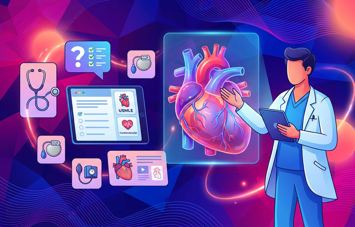
USMLE cardiovascular anatomy focuses on the structural organization of the heart, its great vessels, and the coronary circulation essential for maintaining systemic and pulmonary blood flow. This field requires a firm grasp of the spatial relationships between cardiac chambers, the conduction system, and the specific vascular territories supplied by the coronary arteries. For medical students, recognizing these structures on imaging or during physical exams is a foundational step in USMLE Prep. The heart is situated within the middle mediastinum, protected by the pericardium, and is characterized by four chambers that work in a coordinated sequence. Key anatomical landmarks include the SA node at the junction of the superior vena cava and right atrium, and the AV node located in the Koch triangle. Understanding these relationships is vital because many clinical presentations, such as myocardial infarctions or valvular diseases, are directly linked to specific anatomical defects or vascular occlusions.
Solved Examples
- Coronary Dominance: A patient is found to have the posterior interventricular artery (posterior descending artery) arising from the right coronary artery. Which term best describes this anatomical arrangement?
- Identify the source of the posterior descending artery (PDA).
- Recall that dominance is determined by which artery gives rise to the PDA.
- Since the PDA arises from the right coronary artery (RCA), the heart is right-dominant. This occurs in approximately 85% of the population.
- Cardiac Borders: During a chest X-ray, the right heart border is noted to be enlarged. Which cardiac chamber primarily forms this border?
- Visualize the heart in an anterior-posterior (AP) view.
- The right border is composed almost entirely of the right atrium.
- The left border is primarily formed by the left ventricle and a small portion of the left auricle.
- Aortic Arch Branches: List the three major branches of the aortic arch in order from proximal to distal.
- Locate the aortic arch as it exits the left ventricle.
- The first branch is the brachiocephalic trunk (which later splits into the right common carotid and right subclavian).
- The second branch is the left common carotid artery.
- The third branch is the left subclavian artery.
Practice Questions
1. Which of the following structures is located most posteriorly in the mediastinum and can be compressed by an enlarged left atrium?
2. A 65-year-old man presents with an acute myocardial infarction. Imaging shows an occlusion in the artery that runs in the anterior interventricular sulcus. Which artery is most likely affected?
3. During a cardiac procedure, a surgeon needs to identify the AV node. In which specific anatomical region of the right atrium is the AV node located?
Ready to elevate your USMLE preparation?
Practice with AI-powered USMLE questions, personalized quizzes, adaptive learning, and detailed explanations.
Start USMLE Prep Free4. The "widow maker" refers to a complete occlusion of the proximal left anterior descending artery. Which part of the cardiac conduction system is most at risk of ischemia in this scenario?
5. A patient presents with a stab wound just to the left of the sternum at the level of the 4th intercostal space. Which cardiac chamber is most likely to be injured first?
6. Which heart valve consists of only two cusps and is the most common site for age-related calcification or congenital defects leading to stenosis?
7. The ligamentum arteriosum is a fibrous remnant of a fetal structure. Between which two major vessels does this ligament connect?
8. Which vessel carries oxygenated blood from the lungs back to the left atrium?
9. A patient is diagnosed with an occlusion of the circumflex artery. Which surface of the heart is primarily supplied by this vessel?
10. What is the anatomical name for the rough, muscular ridges found within the right and left auricles and the anterior part of the right atrium?
Answers & Explanations
- Esophagus: The left atrium is the most posterior chamber of the heart. When it enlarges (e.g., due to mitral stenosis), it can compress the esophagus, leading to dysphagia (difficulty swallowing).
- Left Anterior Descending (LAD) Artery: The LAD travels within the anterior interventricular sulcus and supplies the anterior two-thirds of the interventricular septum and the apex.
- Koch Triangle: The AV node is located in the endocardium of the right atrium, specifically within the Koch triangle, bounded by the tendon of Todaro, the tricuspid valve annulus, and the coronary sinus orifice.
- Bundle Branches: The LAD provides the primary blood supply to the anterior portion of the interventricular septum, where the Right Bundle Branch and the Left Anterior Fascicle of the conduction system reside.
- Right Ventricle: The right ventricle forms the majority of the anterior surface of the heart, making it the most vulnerable chamber to penetrating trauma to the anterior chest wall.
- Mitral Valve: The mitral (bicuspid) valve is the only heart valve with two cusps. While the aortic valve is often the site of calcific stenosis, the question specifies the "only two cusps" feature.
- Aorta and Pulmonary Artery: The ligamentum arteriosum is the remnant of the ductus arteriosus, which shunts blood from the pulmonary artery to the aorta in the fetus.
- Pulmonary Veins: Unlike most veins, the four pulmonary veins carry oxygenated blood from the lungs to the left atrium.
- Lateral Wall: The circumflex artery branches off the left main coronary artery and supplies the lateral and posterior walls of the left ventricle.
- Pectinate Muscles: These are the muscular ridges found in the atria. In contrast, the ridges in the ventricles are called trabeculae carneae.
1. Which coronary artery branch typically supplies the SA node in the majority of individuals?
Frequently Asked Questions
What is the most common site of coronary artery occlusion?
The left anterior descending (LAD) artery is the most frequently occluded vessel in myocardial infarctions. It is often referred to as the "widow maker" because it supplies a large portion of the left ventricle.
Where is the best place to auscultate the aortic valve?
The aortic valve is best heard at the second intercostal space at the right sternal border. This location allows for the clearest transmission of sound as blood flows through the valve into the ascending aorta.
What are the layers of the heart wall from outer to inner?
The heart wall consists of the epicardium (visceral layer of serous pericardium), the myocardium (the thick muscular layer), and the endocardium (the innermost endothelial lining). For more on tissue structures, see USMLE Anatomy Practice Questions with Answers.
Which nerve provides parasympathetic innervation to the heart?
The vagus nerve (Cranial Nerve X) provides parasympathetic innervation to the heart, primarily via the cardiac plexus. It acts to slow the heart rate and decrease the force of contraction.
What defines the base of the heart?
The base of the heart is its posterior aspect, formed mainly by the left atrium and a small part of the right atrium. It faces the bodies of thoracic vertebrae T6 through T9.
Ready to elevate your USMLE preparation?
Practice with AI-powered USMLE questions, personalized quizzes, adaptive learning, and detailed explanations.
Start USMLE Prep FreeTags
Enjoyed this article?
Share it with others who might find it helpful.
Related Articles

Hard USMLE Cardiovascular Anatomy Practice Questions
Hard USMLE Cardiovascular Anatomy Practice Questions
Jun 9, 2026
Medium USMLE Cardiovascular Anatomy Practice Questions
Master cardiovascular anatomy for the USMLE Step 1 with these medium-difficulty practice questions, detailed explanations, and high-yield solved examples.
Jun 8, 2026
USMLE Cardiovascular Anatomy Practice Questions with Answers
Master USMLE Cardiovascular Anatomy with these high-yield practice questions, detailed explanations, and clinical pearls for Step 1 preparation.
Jun 8, 2026

Hard USMLE Cardiovascular Physiology Practice Questions
Hard USMLE Cardiovascular Physiology Practice Questions
Jun 9, 2026
Hard USMLE Gastrointestinal Anatomy Practice Questions
Hard USMLE Gastrointestinal Anatomy Practice Questions
Jun 9, 2026
Hard USMLE Musculoskeletal Anatomy Practice Questions
Hard USMLE Musculoskeletal Anatomy Practice Questions
Jun 9, 2026

Hard USMLE Gross Anatomy Practice Questions
Hard USMLE Gross Anatomy Practice Questions
Jun 9, 2026
Hard USMLE Anatomy Practice Questions
Hard USMLE Anatomy Practice Questions
Jun 9, 2026