Hard Physiology Practice Questions Practice Questions

Concept Explanation
Physiology is the scientific study of the functions and mechanisms that work within a living system, encompassing how organisms, organ systems, organs, cells, and biomolecules carry out the chemical and physical functions that exist in a living system. Hard physiology practice questions delve into complex interactions, regulatory feedback loops, and quantitative analysis of physiological processes, often requiring integration of knowledge across multiple organ systems and a deep understanding of underlying cellular and molecular mechanisms. These questions challenge students to apply theoretical knowledge to intricate scenarios, analyze experimental data, and predict outcomes under varying physiological conditions, moving beyond simple recall to true critical thinking.
Solved Examples
Example 1: Renal Physiology
A patient presents with severe dehydration. Their blood osmolarity is 310 mOsm/L (normal: 285-295 mOsm/L) and blood pressure is 85/50 mmHg (normal: 120/80 mmHg). Discuss the expected hormonal and renal responses to restore fluid balance and blood pressure, specifically focusing on ADH, Aldosterone, and the Renin-Angiotensin-Aldosterone System (RAAS).
- Identify the primary imbalances: The patient has hyperosmolarity (due to dehydration) and hypotension.
- ADH response: The hyperosmolarity will stimulate osmoreceptors in the hypothalamus, leading to increased release of Antidiuretic Hormone (ADH) from the posterior pituitary. ADH acts on the collecting ducts and distal convoluted tubules in the kidneys, increasing their permeability to water via aquaporin insertion, thus promoting water reabsorption and concentrating the urine to conserve body water.
- RAAS activation: The hypotension and decreased renal perfusion pressure will stimulate juxtaglomerular cells in the kidneys to release renin. Renin converts angiotensinogen to angiotensin I, which is then converted to angiotensin II by Angiotensin-Converting Enzyme (ACE).
- Angiotensin II effects: Angiotensin II is a potent vasoconstrictor, which will help increase systemic blood pressure. It also stimulates the adrenal cortex to release aldosterone.
- Aldosterone response: Aldosterone acts on the principal cells of the collecting ducts and distal convoluted tubules, increasing sodium reabsorption and potassium secretion. Water follows sodium, thus increasing water reabsorption and contributing to increased blood volume and blood pressure. Aldosterone also directly stimulates ADH release and thirst.
- Overall effect: The combined actions of ADH, Angiotensin II, and Aldosterone work synergistically to increase water reabsorption, retain sodium, raise blood pressure, and stimulate thirst, all aimed at restoring fluid balance and blood pressure.
Example 2: Cardiovascular Regulation
A highly trained endurance athlete has a resting heart rate of 45 bpm and a stroke volume of 120 mL/beat. A sedentary individual has a resting heart rate of 70 bpm and a stroke volume of 70 mL/beat. Both have a mean arterial pressure (MAP) of 90 mmHg. Compare and contrast their cardiac output and total peripheral resistance (TPR) at rest, and explain the physiological adaptations contributing to the athlete's values.
- Calculate Cardiac Output (CO) for both individuals:
- Athlete CO = HR x SV = 45 bpm x 120 mL/beat = 5400 mL/min = 5.4 L/min
- Sedentary CO = HR x SV = 70 bpm x 70 mL/beat = 4900 mL/min = 4.9 L/min
- Calculate Total Peripheral Resistance (TPR) for both individuals:
- TPR = MAP / CO (assuming CVP is negligible)
- Athlete TPR = 90 mmHg / 5.4 L/min ≈ 16.67 mmHg/(L/min)
- Sedentary TPR = 90 mmHg / 4.9 L/min ≈ 18.37 mmHg/(L/min)
- Compare and Contrast: The athlete has a higher cardiac output despite a significantly lower heart rate, due to a much larger stroke volume. The athlete also has a lower total peripheral resistance compared to the sedentary individual, indicating more efficient blood flow through their vascular system.
- Explain Athlete's Adaptations: The athlete's lower resting heart rate is due to increased parasympathetic (vagal) tone and intrinsic cardiac adaptations. The higher stroke volume is a result of physiological hypertrophy of the left ventricle, leading to increased end-diastolic volume (preload) and enhanced contractility. The lower TPR is often attributed to increased capillarization in muscles, greater arterial elasticity, and potentially more efficient endothelial function, allowing for better vasodilation.
Example 3: Respiratory Physiology
A mountaineer ascends to an altitude where the partial pressure of oxygen (PO2) is significantly reduced. Describe the immediate and long-term physiological adaptations in their respiratory and cardiovascular systems to counteract the effects of hypoxia.
- Immediate Respiratory Response: The decreased arterial PO2 is detected by peripheral chemoreceptors (carotid and aortic bodies), leading to an increase in respiratory rate and tidal volume (hyperventilation). This increases alveolar ventilation, helping to raise alveolar PO2 and thus arterial PO2, albeit at the cost of blowing off more CO2, leading to respiratory alkalosis.
- Immediate Cardiovascular Response: Hypoxia also triggers an increase in heart rate and cardiac output to enhance oxygen delivery to tissues. Pulmonary vasoconstriction occurs in response to localized alveolar hypoxia, shunting blood away from poorly oxygenated alveoli to better-ventilated ones.
- Long-term Hematological Adaptation: Chronic hypoxia stimulates the kidneys to release erythropoietin (EPO). EPO stimulates the bone marrow to produce more red blood cells (erythrocytosis), increasing the hematocrit and oxygen-carrying capacity of the blood.
- Long-term Respiratory Adaptation: The respiratory alkalosis initially inhibits ventilation, but over days, the kidneys compensate by excreting bicarbonate, allowing ventilation to increase further without severe pH disturbances. Increased efficiency of oxygen utilization by tissues also occurs.
- Long-term Cardiovascular Adaptation: While cardiac output increases initially, chronic altitude exposure can lead to pulmonary hypertension due to widespread pulmonary vasoconstriction and vascular remodeling. Systemic vascular resistance may decrease slightly due to generalized vasodilation in systemic tissues to improve oxygen delivery.
Practice Questions
1. A patient with uncontrolled type 1 diabetes mellitus presents with severe metabolic acidosis (blood pH 7.15, normal 7.35-7.45) due to diabetic ketoacidosis. Explain the primary compensatory mechanism the body will employ to normalize blood pH, detailing the specific physiological changes that occur.
2. Discuss the precise roles of the sarcoplasmic reticulum, T-tubules, and calcium ions in skeletal muscle contraction, from the arrival of an action potential at the neuromuscular junction to the initiation of muscle fiber shortening.
3. A novel drug is developed that selectively inhibits the activity of voltage-gated Na+ channels in neurons. Predict the effect of this drug on the generation and propagation of action potentials, and explain the consequences for synaptic transmission.
4. Explain how the countercurrent multiplier system in the loop of Henle, in conjunction with the countercurrent exchange system of the vasa recta, enables the kidney to produce concentrated urine. Quantify the osmotic gradient created and maintained.
5. A patient is diagnosed with a deficiency in pancreatic lipase. Describe the specific impact this will have on the digestion and absorption of dietary fats and fat-soluble vitamins, tracing the pathway from the small intestine to systemic circulation.
6. Detail the process of excitation-contraction coupling in cardiac muscle, highlighting the key differences compared to skeletal muscle, particularly regarding the source and role of calcium ions and the duration of the action potential.
7. A patient experiences a sudden drop in blood glucose levels. Explain the hormonal cascade initiated to restore glucose homeostasis, focusing on the roles of specific pancreatic hormones and their target tissues.
8. Describe the intricate feedback mechanisms involved in the regulation of thyroid hormone synthesis and release, starting from the hypothalamus and detailing the effects of both negative and positive feedback loops.
Answers & Explanations
1. Answer: The primary compensatory mechanism for metabolic acidosis is respiratory compensation, specifically an increase in alveolar ventilation. The severe metabolic acidosis (low pH) will be detected by peripheral chemoreceptors (carotid and aortic bodies) and central chemoreceptors (in the medulla). These chemoreceptors stimulate the respiratory center in the brainstem, leading to an increased respiratory rate and depth (hyperventilation). This increased ventilation expels more CO2 from the body, lowering arterial PCO2. According to the Henderson-Hasselbalch equation (pH = pKa + log([HCO3-]/[PCO2])), a decrease in PCO2 will increase the pH, thereby buffering the metabolic acid load and helping to restore blood pH towards normal. This is a rapid but partial compensation. Renal compensation, which involves increased acid excretion and bicarbonate reabsorption/generation, would also occur but is slower.
2. Answer: Upon arrival of an action potential at the neuromuscular junction, acetylcholine is released, binding to receptors on the sarcolemma and generating an end-plate potential that triggers an action potential in the muscle fiber. This action potential propagates along the sarcolemma and dives deep into the muscle fiber via invaginations called T-tubules (transverse tubules). As the action potential travels down the T-tubules, it causes a conformational change in dihydropyridine receptors (DHPRs), which are voltage-sensitive proteins. These DHPRs are mechanically linked to ryanodine receptors (RyRs) on the adjacent sarcoplasmic reticulum (SR). The conformational change in DHPRs physically pulls open the RyRs, which are calcium release channels on the SR. This causes a massive efflux of stored calcium ions (Ca2+) from the SR into the sarcoplasm. These Ca2+ ions then bind to troponin C on the thin filaments, initiating the cross-bridge cycle and muscle contraction by moving tropomyosin away from actin binding sites, allowing myosin heads to attach to actin.
3. Answer: If a drug selectively inhibits voltage-gated Na+ channels, the generation of action potentials will be severely impaired or completely blocked. When a neuron receives a sufficient depolarizing stimulus, these channels are crucial for the rapid influx of Na+ ions that causes the rapid rising phase (depolarization) of the action potential. Without functional voltage-gated Na+ channels, the threshold potential cannot be reached, or if it is, the depolarization will be insufficient to propagate a full action potential. Consequently, action potentials will not be generated at the axon hillock and thus cannot be propagated along the axon. This lack of action potential propagation means that no electrical signal will reach the axon terminals. Therefore, the release of neurotransmitters from the presynaptic terminal will cease, leading to a complete disruption of synaptic transmission and communication between neurons. This would result in widespread neurological dysfunction, affecting sensory perception, motor control, and cognitive processes.
4. Answer: The countercurrent multiplier system in the loop of Henle establishes a vertical osmotic gradient in the renal medulla, while the countercurrent exchange system of the vasa recta maintains it. In the descending limb of the loop of Henle, water is passively reabsorbed due to the increasing medullary tonicity, making the tubular fluid more concentrated. The ascending limb is impermeable to water but actively transports Na+, K+, and Cl- out of the tubule into the interstitial fluid, further increasing medullary osmolarity. This active transport is the 'multiplier' effect, as it continuously adds solutes to the medulla, creating an osmotic gradient that can reach up to 1200 mOsm/L at the deepest part of the medulla (from ~300 mOsm/L in the cortex). The vasa recta, capillaries running parallel to the loop of Henle, act as countercurrent exchangers. As blood flows down into the medulla, it picks up solutes and loses water, becoming more concentrated. As it flows back up, it loses solutes and picks up water. This arrangement minimizes the washout of solutes from the medullary interstitium, preserving the osmotic gradient. The collecting ducts then pass through this osmotic gradient; in the presence of ADH, water is reabsorbed from the collecting ducts into the hypertonic medulla, resulting in concentrated urine.
5. Answer: A deficiency in pancreatic lipase will significantly impair the digestion and subsequent absorption of dietary fats (triglycerides) and fat-soluble vitamins (A, D, E, K). Pancreatic lipase is the primary enzyme responsible for breaking down triglycerides into monoglycerides and free fatty acids in the small intestine. Without sufficient lipase, fats will not be properly emulsified and hydrolyzed, leading to a condition called steatorrhea (excess fat in feces). This undigested fat will also impair the formation of micelles, which are crucial for solubilizing monoglycerides, free fatty acids, and fat-soluble vitamins to allow their transport to the brush border of enterocytes for absorption. Consequently, absorption of these essential nutrients will be drastically reduced. The unabsorbed fats and vitamins will pass into the large intestine, causing gastrointestinal symptoms such as bloating, abdominal pain, and foul-smelling stools. Over time, this malabsorption can lead to deficiencies in fat-soluble vitamins, impacting vision (Vit A), bone health (Vit D), antioxidant protection (Vit E), and blood clotting (Vit K).
6. Answer: Excitation-contraction coupling in cardiac muscle begins with an action potential propagating through gap junctions to adjacent cardiac myocytes. Unlike skeletal muscle, the cardiac action potential has a prolonged plateau phase, primarily due to the sustained influx of Ca2+ through voltage-gated L-type calcium channels (DHP receptors) in the sarcolemma and T-tubules. This extracellular Ca2+ influx is crucial; it triggers the release of a much larger amount of Ca2+ from the sarcoplasmic reticulum (SR) via ryanodine receptors (RyRs) in a process called calcium-induced calcium release (CICR). This initial influx of extracellular Ca2+ is therefore essential for stimulating the main SR Ca2+ release, acting as a 'trigger calcium.' The released Ca2+ then binds to troponin C, initiating the cross-bridge cycle and muscle contraction, similar to skeletal muscle. Key differences from skeletal muscle include: 1) the reliance on extracellular Ca2+ influx to trigger SR release, 2) the longer duration of the action potential (due to the Ca2+ plateau) which prevents tetany and ensures adequate filling time for the heart, and 3) the presence of gap junctions for electrical coupling, allowing the heart to contract as a functional syncytium.
7. Answer: A sudden drop in blood glucose (hypoglycemia) primarily triggers a hormonal cascade involving the pancreas and adrenal glands to restore glucose homeostasis. The primary response comes from the alpha cells of the pancreatic islets, which detect the low glucose and secrete glucagon. Glucagon acts primarily on the liver, stimulating glycogenolysis (breakdown of stored glycogen into glucose) and gluconeogenesis (synthesis of new glucose from non-carbohydrate precursors like amino acids and glycerol). This releases glucose into the bloodstream, raising blood glucose levels. Additionally, the adrenal medulla releases epinephrine (and norepinephrine) in response to hypoglycemia, which also promotes hepatic glycogenolysis and gluconeogenesis, and inhibits insulin secretion. Over a longer term, cortisol from the adrenal cortex also contributes to gluconeogenesis. The combined actions of these hormones ensure rapid and sustained elevation of blood glucose to prevent cellular energy deficit, especially in the brain.
8. Answer: The regulation of thyroid hormone (TH) synthesis and release is governed by a classic negative feedback loop involving the hypothalamus, anterior pituitary, and thyroid gland, often referred to as the Hypothalamic-Pituitary-Thyroid (HPT) axis. The process begins in the hypothalamus, which secretes Thyrotropin-Releasing Hormone (TRH). TRH travels through the hypothalamo-hypophyseal portal system to the anterior pituitary, stimulating the release of Thyroid-Stimulating Hormone (TSH, also known as thyrotropin). TSH then acts on the thyroid gland, promoting all steps of TH synthesis and release, including iodine uptake, thyroglobulin synthesis, follicular cell growth, and release of primarily thyroxine (T4) and smaller amounts of triiodothyronine (T3). Once released, T3 and T4 exert negative feedback. High levels of T3 and T4 inhibit TRH release from the hypothalamus and TSH release from the anterior pituitary, thereby reducing further stimulation of the thyroid gland. This negative feedback loop ensures that thyroid hormone levels are maintained within a narrow physiological range. There are no significant positive feedback loops in the normal regulation of thyroid hormones; however, certain pathologies or pharmacological interventions can disrupt this delicate balance.
1. Which of the following is the primary compensatory mechanism for metabolic acidosis?
Frequently Asked Questions
What is the difference between positive and negative feedback in physiology?
Negative feedback loops are much more common and work to stabilize a physiological variable by counteracting a change, bringing the system back to its set point. Positive feedback loops amplify a change, moving the system further away from its set point, and typically require an external event to terminate the process, such as childbirth or blood clotting.
How do the kidneys regulate blood pressure?
The kidneys regulate blood pressure through several mechanisms: controlling blood volume by adjusting water and sodium excretion, releasing renin to activate the Renin-Angiotensin-Aldosterone System (RAAS) which affects vasoconstriction and fluid retention, and producing other vasoactive substances. These actions collectively influence cardiac output and total peripheral resistance.
Why is oxygen delivery so critical for physiological function?
Oxygen is the final electron acceptor in the electron transport chain, which is the primary pathway for ATP production (cellular energy) via oxidative phosphorylation. Without sufficient oxygen, cells cannot produce enough ATP to sustain vital functions, leading to cellular dysfunction and death, particularly in highly metabolic organs like the brain and heart.
What is the role of the autonomic nervous system in regulating organ function?
The autonomic nervous system (ANS), comprising the sympathetic and parasympathetic divisions, involuntarily regulates the function of internal organs. The sympathetic system generally prepares the body for 'fight or flight' responses, while the parasympathetic system promotes 'rest and digest' activities, maintaining homeostasis through opposing actions on target organs like the heart, lungs, and digestive tract. For more on the nervous system, check out our Nervous System Questions Practice Questions.
How does the body maintain fluid balance?
The body maintains fluid balance through a complex interplay of hormonal and neural mechanisms, primarily involving the kidneys, hypothalamus, and cardiovascular system. Key hormones include Antidiuretic Hormone (ADH) which regulates water reabsorption, and Aldosterone which controls sodium reabsorption, both influencing blood volume and osmolarity. Thirst mechanisms also play a crucial role in water intake.
What is the significance of the Frank-Starling law of the heart?
The Frank-Starling law states that the stroke volume of the heart increases in response to an increase in the volume of blood filling the heart (end-diastolic volume), when all other factors remain constant. This intrinsic mechanism ensures that the heart ejects all the blood returned to it, maintaining balance between venous return and cardiac output, and preventing blood from pooling in the venous circulation. For more cardiovascular insights, see our Cardiovascular System Questions Practice Questions.
Enjoyed this article?
Share it with others who might find it helpful.
Related Articles

Medium Physiology Practice Questions Practice Questions
Boost your understanding of physiological processes with these medium physiology practice questions covering homeostasis, organ systems, and cellular functions.
Apr 25, 2026

Easy Physiology Practice Questions Practice Questions
These easy physiology practice questions cover fundamental concepts of how the human body functions, including organ systems, cells, and homeostatic mechanisms.
Apr 25, 2026
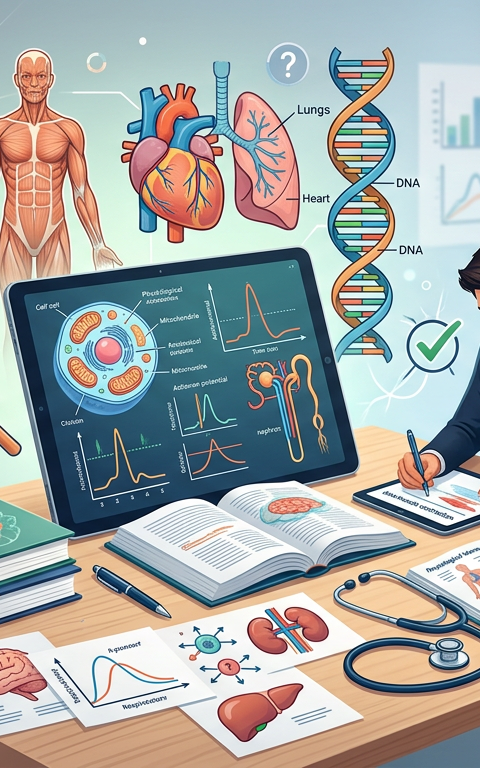
Physiology Practice Questions Practice Questions with Answers
Dive into comprehensive Physiology Practice Questions covering homeostasis, organ systems, and cellular functions with detailed explanations and a quick quiz.
Apr 25, 2026
Hard Microbiology Practice Questions Practice Questions
Hard Microbiology Practice Questions Practice Questions
Apr 26, 2026

Medium Microbiology Practice Questions Practice Questions
Master microbiology with our medium-level practice questions covering bacterial growth, genetics, and metabolism. Includes solved examples and a quick quiz.
Apr 26, 2026

Easy Microbiology Practice Questions Practice Questions
Easy Microbiology Practice Questions Practice Questions
Apr 26, 2026

Hard Enzyme Questions Practice Questions
Master complex biochemistry with these hard enzyme questions. Cover Michaelis-Menten kinetics, inhibition types, and Lineweaver-Burk plots with solved examples.
Apr 26, 2026

Medium Enzyme Questions Practice Questions
Master enzyme kinetics and regulation with our Medium Enzyme Questions Practice Questions. Includes solved examples, detailed explanations, and a quick quiz.
Apr 26, 2026