Hard Cell Structure Practice Questions Practice Questions
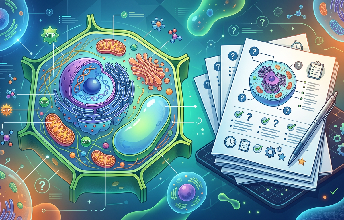
Hard Cell Structure Practice Questions Practice Questions
Mastering cell biology requires moving beyond simple memorization of organelles and their functions. To truly understand the cell, you must analyze it as a dynamic, interconnected system. This page provides hard cell structure practice questions designed to test your ability to synthesize information, predict outcomes of cellular disruptions, and apply your knowledge to complex biological scenarios. These problems will challenge you to think critically about how the intricate architecture of the cell dictates its ability to live, grow, and respond to its environment.
Concept Explanation
Advanced cell structure refers to the intricate, dynamic, and interconnected network of organelles and macromolecules that perform complex life-sustaining functions within a eukaryotic cell. This concept moves beyond identifying organelles to understanding how they work together in complex pathways like the endomembrane system, how the cytoskeleton provides a dynamic framework for transport and division, and how energy-converting organelles like mitochondria power cellular activities. What makes this topic hard is the focus on the system's integration; a defect in one component, such as a single protein in the endoplasmic reticulum, can have cascading effects on protein secretion, membrane integrity, and overall cell viability. Understanding hard cell structure involves analyzing the cell not as a static diagram, but as a bustling city with coordinated traffic, energy production, waste management, and communication systems, all governed by the laws of chemistry and physics.
Solved Examples of Hard Cell Structure Questions
The following examples demonstrate how to approach complex problems related to cell structure and function. Each solution breaks down the problem into logical steps, connecting a given scenario to its underlying cellular mechanisms.
Example 1: Mitochondrial Dysfunction
Question: A patient is diagnosed with a rare genetic disorder caused by a mutation in a gene encoding a protein subunit of Complex I (NADH dehydrogenase) in the mitochondrial electron transport chain. Explain the primary and secondary cellular consequences of this mutation, particularly concerning ATP production and the cell's metabolic state.
Solution:
- Identify the primary defect: The mutation affects Complex I of the electron transport chain (ETC). This complex is responsible for accepting electrons from NADH and pumping protons (H+) from the mitochondrial matrix into the intermembrane space.
- Analyze the immediate impact on the ETC: A non-functional Complex I means that NADH cannot effectively donate its electrons to the ETC. This severely impairs the flow of electrons through the chain to oxygen.
- Determine the effect on the proton gradient: Since Complex I is a major proton pump, its failure reduces the number of protons pumped into the intermembrane space. This weakens the proton-motive force, which is the electrochemical gradient that powers ATP synthase.
- Explain the impact on ATP synthesis: With a diminished proton-motive force, ATP synthase cannot produce ATP at a normal rate. This leads to a severe energy deficit in the cell, as oxidative phosphorylation is the most efficient method of ATP production.
- Consider the secondary metabolic shifts: To compensate for the lack of ATP from oxidative phosphorylation, the cell will heavily increase its reliance on glycolysis. This leads to the rapid conversion of glucose to pyruvate. Because the mitochondria cannot efficiently process the pyruvate and the resulting NADH, the cell will convert pyruvate to lactate (in animals) to regenerate NAD+ for glycolysis to continue. This results in lactic acidosis, a buildup of lactic acid in tissues and the bloodstream, causing fatigue and muscle pain.
Example 2: Cytoskeletal Disruption
Question: A researcher treats a culture of rapidly dividing epithelial cells with Taxol, a drug that binds to and stabilizes microtubules, preventing their depolymerization. Describe the most significant effect on the cell cycle and explain why.
Solution:
- Identify the target: Taxol targets microtubules, a key component of the cytoskeleton. Specifically, it prevents their disassembly (depolymerization).
- Relate microtubules to the cell cycle: Microtubules form the mitotic spindle during M-phase (mitosis). The spindle is responsible for capturing chromosomes at their kinetochores and then segregating the sister chromatids to opposite poles of the cell.
- Analyze the function of microtubule dynamics in mitosis: The process of chromosome segregation is highly dynamic. Kinetochore microtubules must be able to shorten (depolymerize) to pull the sister chromatids apart during anaphase.
- Predict the effect of Taxol: By stabilizing microtubules and preventing them from shortening, Taxol effectively freezes the mitotic spindle. The cell can form a spindle and align chromosomes on the metaphase plate, but it cannot progress to anaphase because the signal to separate sister chromatids is linked to the tension provided by dynamic microtubules. The spindle assembly checkpoint will remain active, arresting the cell in metaphase.
- State the conclusion: The most significant effect is cell cycle arrest in metaphase. The cell is unable to complete mitosis, which ultimately triggers apoptosis (programmed cell death). This is the mechanism by which Taxol functions as a chemotherapy agent against cancer.
Example 3: Protein Trafficking Error
Question: A cell is genetically engineered to produce a lysosomal hydrolase, but the gene has been altered so that the mannose-6-phosphate (M6P) tag is not added to the protein in the cis-Golgi. Where will this protein end up, and what will be the consequence for the cell?
Solution:
- Trace the normal pathway: A lysosomal hydrolase is synthesized in the rough ER, moves to the Golgi apparatus, and then is sorted to the lysosome.
- Identify the sorting signal: The key sorting signal that directs proteins from the trans-Golgi network to the lysosome is the mannose-6-phosphate (M6P) tag. This tag is recognized by M6P receptors in the trans-Golgi, which package the hydrolases into vesicles destined for the lysosome.
- Analyze the effect of the missing signal: Without the M6P tag, the Golgi apparatus does not recognize the hydrolase as a lysosomal protein. The cell's protein sorting machinery will treat it as a default secretory protein.
- Determine the final destination: The protein will be packaged into a secretory vesicle at the trans-Golgi network. This vesicle will travel to the plasma membrane, fuse with it, and release the hydrolase outside the cell via exocytosis. This is known as the default secretory pathway. For more information on cellular pathways, you can explore resources like the Scitable by Nature Education.
- Describe the cellular consequence: The cell's lysosomes will lack this specific hydrolase. If this is a critical enzyme, its substrate will accumulate within the lysosomes, leading to swelling of the organelles and the development of a lysosomal storage disease, similar to I-cell disease.
Practice Questions
Test your understanding with these hard cell structure practice questions.
1. (Easy) A cell actively secretes a large volume of protein-based hormones. Which two organelles would you expect to be exceptionally abundant and metabolically active in this cell? Explain your reasoning.
2. (Medium) A toxin is introduced that specifically and irreversibly blocks the channel of aquaporins in the cell membrane. How would this affect a red blood cell if it were placed in a hypotonic solution? Compare this to a normal red blood cell under the same conditions.
3. (Hard) You are studying a cell with a mutation that results in a dysfunctional signal recognition particle (SRP). Describe the immediate consequences for the synthesis and final location of (a) a cytoplasmic enzyme like phosphofructokinase and (b) a secreted protein like collagen.
4. (Medium) Compare the primary functions of the smooth endoplasmic reticulum (SER) and the peroxisome. Provide one function they share and two distinct functions for each.
5. (Hard) Dinitrophenol (DNP) is a chemical that acts as a protonophore, meaning it can transport protons across the inner mitochondrial membrane, bypassing ATP synthase. If a cell is exposed to DNP, what happens to (a) the proton gradient, (b) the rate of ATP synthesis by oxidative phosphorylation, and (c) the rate of oxygen consumption? Explain your reasoning for each.
6. (Hard) Design a simple experiment using fluorescence microscopy to prove that the Golgi apparatus, and not the endoplasmic reticulum, is responsible for sorting proteins to different cellular locations. Assume you can tag your protein of interest with Green Fluorescent Protein (GFP) and can apply drugs that block transport between organelles.
7. (Medium) Explain the structural basis of the endosymbiotic theory. What specific features of mitochondria and chloroplasts are cited as evidence for this theory?
8. (Hard) A cell has a defect in the enzyme that builds the oligosaccharide precursor in the rough ER lumen (part of N-linked glycosylation). How would this defect impact the folding and quality control of glycoproteins within the ER? How might this affect the cell's ability to interact with its environment? Advanced techniques like NMR interpretation are often used to study the structure of these complex molecules.
Answers & Explanations
1. Answer: The rough endoplasmic reticulum (RER) and the Golgi apparatus.
Explanation: Protein-based hormones are synthesized for secretion. This process begins on ribosomes attached to the RER, where the polypeptide chain is fed into the ER lumen for folding and modification. From the RER, the proteins are transported in vesicles to the Golgi apparatus for further processing, sorting, and packaging into secretory vesicles. Therefore, a cell specialized for protein secretion, like a pancreatic islet cell secreting insulin, would have a vast RER and a large, complex Golgi.
2. Answer: A normal red blood cell in a hypotonic solution would rapidly swell and burst (hemolysis) due to the fast influx of water through aquaporins. The cell with blocked aquaporins would still swell and eventually burst, but at a much slower rate.
Explanation: Osmosis is the movement of water across a semipermeable membrane from an area of low solute concentration (hypotonic solution) to high solute concentration (the cytoplasm). While water can diffuse slowly across the lipid bilayer, aquaporins are specialized channels that facilitate rapid water transport. In a normal cell, the osmotic gradient causes a massive, rapid influx of water through these channels, leading to lysis. In the cell with blocked aquaporins, water can only enter via slow diffusion across the membrane, delaying the swelling and lysis.
3. Answer: (a) The synthesis and location of phosphofructokinase would be unaffected. (b) The synthesis of collagen would complete in the cytoplasm, and it would remain there instead of being secreted.
Explanation: The Signal Recognition Particle (SRP) is responsible for identifying the ER signal sequence on a newly forming polypeptide chain and directing the entire ribosome-mRNA-polypeptide complex to the RER.
(a) Phosphofructokinase is a cytoplasmic enzyme; its mRNA is translated on free ribosomes in the cytoplasm and it lacks an ER signal sequence. The SRP is not involved in its synthesis, so a dysfunctional SRP has no effect.
(b) Collagen is a secreted protein and thus must enter the endomembrane system. Its synthesis begins on a free ribosome, and an ER signal sequence emerges. Normally, the SRP would bind this sequence and pause translation until the complex docks at the RER. With a dysfunctional SRP, the signal sequence is not recognized. Translation continues and completes on the free ribosome, releasing the full collagen protein into the cytoplasm, where it cannot be folded, modified, or secreted correctly.
4. Answer: Both the SER and peroxisomes are involved in aspects of lipid metabolism. Distinct functions for the SER include calcium storage and detoxification of drugs/poisons. Distinct functions for the peroxisome include breaking down fatty acids via beta-oxidation and neutralizing toxic substances like hydrogen peroxide.
Explanation:
Shared Function: Both organelles participate in lipid metabolism. The SER is the primary site of synthesis for lipids like steroids and phospholipids. Peroxisomes break down very long-chain fatty acids.
Distinct SER Functions: It sequesters and releases calcium ions (Ca2+), a critical function for cell signaling, especially in muscle cells (where it's called the sarcoplasmic reticulum). It also contains enzymes that detoxify drugs and poisons, often by adding hydroxyl groups to make them more water-soluble.
Distinct Peroxisome Functions: They contain oxidases that use oxygen to break down molecules, producing hydrogen peroxide (H2O2) as a byproduct. They also contain the enzyme catalase, which breaks down the toxic H2O2 into water and oxygen.
5. Answer: (a) The proton gradient will be dissipated. (b) The rate of ATP synthesis will drop to nearly zero. (c) The rate of oxygen consumption will increase.
Explanation: DNP uncouples oxidative phosphorylation.
(a) By providing an alternate route for protons to flow back into the matrix, DNP eliminates the proton-motive force. The gradient cannot be maintained.
(b) ATP synthase relies entirely on the flow of protons through its channel to generate ATP. Since DNP dissipates this gradient, ATP synthesis via this mechanism halts.
(c) The electron transport chain (ETC) and ATP synthesis are coupled; a high concentration of ATP or a strong proton gradient can inhibit the ETC (feedback inhibition). By dissipating the gradient, DNP removes this inhibition. The ETC works at its maximum rate, consuming NADH and FADH2 and passing electrons to oxygen, the final electron acceptor. Thus, oxygen consumption increases dramatically, but the energy is released as heat instead of being captured in ATP.
6. Answer: The experiment would involve a 'pulse-chase' design with a temperature-sensitive transport block.
Explanation:
1. Construct: Engineer cells to express a GFP-tagged protein that has two different sorting signals: one for the peroxisome and one for secretion.
2. Pulse: Synthesize a burst of this protein. All synthesis will occur in the RER. Initially, GFP fluorescence will be seen exclusively in the ER network.
3. ER-to-Golgi Transport: Allow the protein to travel from the ER to the Golgi apparatus. You can confirm its arrival by colocalization with a Golgi-specific marker (e.g., a red fluorescent protein tagged to a Golgi-resident enzyme).
4. The Block: Apply a drug like Brefeldin A, which causes the Golgi to collapse back into the ER, or use a temperature block (e.g., 20°C) that allows proteins to enter the Golgi but not leave the trans-Golgi network. This traps the protein in the Golgi.
5. Chase and Observation: Wash out the drug or change the temperature to allow transport to resume. Observe the GFP signal. You would see one population of GFP moving to small vesicles that travel to the cell periphery and fuse with the membrane (secretion), while another population moves to different vesicles that target the peroxisomes (identified by another fluorescent marker). Because the protein was held in a single location (the Golgi) before splitting into two distinct paths, this proves the Golgi is the sorting station. If the ER were the sorter, you would have seen two different types of transport vesicles budding directly from the ER, which is not observed.
7. Answer: The endosymbiotic theory proposes that mitochondria and chloroplasts originated as free-living prokaryotic cells that were engulfed by an ancestral eukaryotic cell. The evidence lies in their unique structural features.
Explanation:
1. Double Membrane: Both organelles have two membranes. The inner membrane is proposed to be the original prokaryotic plasma membrane, while the outer membrane is derived from the host cell's vesicle membrane during engulfment. The inner membrane has proteins (like ETC components) different from other eukaryotic membranes.
2. Own DNA: They contain their own single, circular DNA molecule, much like the chromosome of a bacterium. This DNA lacks histones found in eukaryotic nuclear DNA.
3. Own Ribosomes: Their ribosomes (70S) are more similar in size and sequence to prokaryotic ribosomes than to the eukaryotic ribosomes (80S) found in the cytoplasm.
4. Replication: They replicate independently of the cell nucleus through a process resembling binary fission, the method of prokaryotic cell division.
For a comprehensive overview, see the UC Berkeley evolution page on endosymbiosis.
8. Answer: The defect would severely impair glycoprotein folding and quality control, likely leading to the accumulation of misfolded proteins and triggering the Unfolded Protein Response (UPR). This would compromise cell surface receptors and cell-cell adhesion molecules.
Explanation: The large oligosaccharide precursor added in N-linked glycosylation acts as a tag for proper folding. Chaperone proteins in the ER, like calnexin and calreticulin, specifically bind to these sugar chains to assist the glycoprotein in achieving its correct three-dimensional shape. This process is complex, and simplifying expressions of these pathways can help in understanding. Without the initial sugar chain, these chaperones cannot bind, and the protein is highly likely to misfold. The ER's quality control system identifies misfolded proteins and attempts to refold them or targets them for degradation (ER-associated degradation, or ERAD). A widespread failure in glycosylation would overwhelm this system, causing misfolded proteins to accumulate and triggering the UPR, which can lead to apoptosis. Since many cell surface receptors, channels, and cell adhesion molecules are glycoproteins, this defect would severely impair the cell's ability to receive signals and physically interact with neighboring cells and the extracellular matrix.
1. A drug that prevents the polymerization of actin filaments is added to a motile cell. Which of the following processes would be most directly inhibited?
Frequently Asked Questions
What is the functional difference between the rough ER and smooth ER?
The primary functional difference lies in their associated enzymes and whether they have ribosomes. The rough ER is studded with ribosomes and is the site of synthesis and modification for secreted proteins, lysosomal proteins, and membrane proteins. The smooth ER lacks ribosomes and is the primary site for lipid synthesis, steroid hormone production, calcium storage, and detoxification.
How do mitochondria and chloroplasts support the theory of endosymbiosis?
Mitochondria and chloroplasts support the endosymbiotic theory because they possess features characteristic of independent prokaryotic organisms. These include a double membrane, their own circular DNA molecule, prokaryote-like 70S ribosomes, and the ability to replicate by a process similar to binary fission, independently of the cell's nuclear division cycle.
Why is the cytoskeleton considered dynamic?
The cytoskeleton is considered dynamic because its primary components—microtubules, actin filaments, and intermediate filaments—are not static structures. They can be rapidly assembled (polymerized) and disassembled (depolymerized), allowing the cell to change shape, move, divide, and rearrange its internal organelles in response to internal and external signals.
What happens if the Golgi apparatus stops working?
If the Golgi apparatus stops working, the cell's ability to process, sort, and transport proteins and lipids would be crippled. Proteins from the ER would not be properly modified or packaged, leading to the failure of secretion, non-functional lysosomes, and incorrect placement of membrane proteins. This systemic failure in cellular logistics would quickly lead to cell death.
How do cells use their structure for communication?
Cells use their structure for communication primarily through the plasma membrane and cell junctions. The plasma membrane contains receptor proteins that bind to external signaling molecules (like hormones), initiating internal signaling cascades. Cell junctions, such as gap junctions (in animals) and plasmodesmata (in plants), form direct cytoplasmic channels between adjacent cells, allowing ions and small molecules to pass through, coordinating their metabolic activities and responses.
Enjoyed this article?
Share it with others who might find it helpful.
Related Articles
Medium Cell Structure Practice Questions Practice Questions
Master cell biology with these medium cell structure practice questions. Get detailed explanations and solved examples to boost your understanding.
Apr 25, 2026
Easy Cell Structure Practice Questions Practice Questions
Master the basics of biology with our easy cell structure practice questions. This guide includes clear explanations, solved examples, and a quiz to test your knowledge.
Apr 25, 2026

Cell Structure Practice Questions Practice Questions with Answers
Cell Structure Practice Questions Practice Questions with Answers
Apr 25, 2026

Hard Microbiology Practice Questions Practice Questions
Hard Microbiology Practice Questions Practice Questions
Apr 26, 2026

Medium Microbiology Practice Questions Practice Questions
Master microbiology with our medium-level practice questions covering bacterial growth, genetics, and metabolism. Includes solved examples and a quick quiz.
Apr 26, 2026

Easy Microbiology Practice Questions Practice Questions
Easy Microbiology Practice Questions Practice Questions
Apr 26, 2026

Hard Enzyme Questions Practice Questions
Master complex biochemistry with these hard enzyme questions. Cover Michaelis-Menten kinetics, inhibition types, and Lineweaver-Burk plots with solved examples.
Apr 26, 2026

Medium Enzyme Questions Practice Questions
Master enzyme kinetics and regulation with our Medium Enzyme Questions Practice Questions. Includes solved examples, detailed explanations, and a quick quiz.
Apr 26, 2026