Hard Anatomy Practice Questions Practice Questions
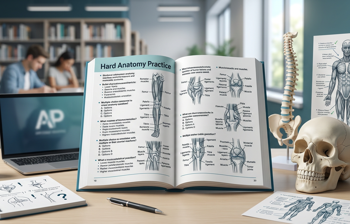
Concept Explanation
Anatomy is the scientific study of the structure of organisms, including their systems, organs, and tissues, and how these components relate to one another spatially and functionally. Mastering Hard Anatomy Practice Questions requires more than just memorizing names; it demands a deep understanding of regional relationships, neurovascular pathways, and the clinical implications of structural variations. Advanced anatomy often bridges the gap between gross morphology and surgical or diagnostic applications. For instance, understanding the cardiovascular system involves not just the heart's chambers, but the precise branching of coronary arteries and the venous drainage patterns that maintain myocardial health. According to resources like Khan Academy, anatomical study is foundational for all medical sciences, providing the map upon which physiology and pathology are written. Whether you are examining the intricate layers of the nervous system or the complex interactions within organ systems, high-level mastery involves predicting how a lesion in one area will manifest as a functional deficit elsewhere.
Solved Examples
- Question: A patient presents with "winging" of the scapula when pushing against a wall. Which nerve is likely damaged, and what is its anatomical origin?
- Identify the primary muscle responsible for holding the scapula against the thoracic wall: the Serratus Anterior.
- Recall the innervation of the Serratus Anterior: the Long Thoracic Nerve.
- Determine the nerve's origin: It arises from the ventral rami of C5, C6, and C7 spinal nerves.
- Conclusion: The Long Thoracic Nerve (C5-C7) is damaged.
- Question: During a thyroidectomy, a surgeon accidentally ligates the artery supplying the superior pole of the thyroid. Which nerve, located in close proximity, is at risk of injury?
- Identify the artery: The Superior Thyroid Artery.
- Recall the neurovascular relationship: The Superior Thyroid Artery travels closely with the External Branch of the Superior Laryngeal Nerve.
- Identify the functional impact: This nerve innervates the cricothyroid muscle, which tenses the vocal folds.
- Conclusion: The External Branch of the Superior Laryngeal Nerve is at risk.
- Question: Trace the flow of cerebrospinal fluid (CSF) from the lateral ventricles to the subarachnoid space.
- CSF is produced in the choroid plexus of the lateral ventricles.
- It flows through the Interventricular Foramen (of Monro) into the Third Ventricle.
- It travels through the Cerebral Aqueduct (of Sylvius) into the Fourth Ventricle.
- It exits the Fourth Ventricle via the Median Aperture (Magendie) and Lateral Apertures (Luschka) to enter the Subarachnoid Space.
Practice Questions
- A 45-year-old male presents with loss of sensation on the skin of the medial aspect of the arm and forearm following a traumatic axillary injury. Which cord of the brachial plexus is likely involved?
- What specific anatomical structure forms the boundary between the upper and lower gastrointestinal tracts, and what is its clinical significance in GI bleeds?
- During a cranial nerve exam, a patient is unable to adduct the left eye during rightward gaze but can adduct the eye during convergence. Where is the most likely site of the lesion?
- Which dural venous sinus lies within the free margin of the falx cerebri and drains into the straight sinus?
- Identify the four muscles of the rotator cuff and specify which one is responsible for the first 15 degrees of arm abduction.
- A patient exhibits "foot drop" and loss of sensation on the dorsal surface of the foot. Which nerve is compressed, and at what bony landmark is it most vulnerable?
- Describe the pathway of the Ophthalmic Artery and identify which major vessel it branches from.
- What are the three components of the portal triad found within the hepatoduodenal ligament?
- Which laryngeal muscle is the only one responsible for abducting the vocal folds, effectively opening the rima glottidis?
- In the cardiac conduction system, what is the anatomical location of the Atrioventricular (AV) node?
Answers & Explanations
- Medial Cord: The medial cord of the brachial plexus gives rise to the medial cutaneous nerve of the arm and the medial cutaneous nerve of the forearm, which provide sensory innervation to those regions.
- Ligament of Treitz: The Suspensory Muscle of the Duodenum (Ligament of Treitz) marks the junction between the duodenum and jejunum. It is the anatomical landmark used to differentiate between Upper GI bleeds (proximal to the ligament) and Lower GI bleeds (distal to the ligament).
- Medial Longitudinal Fasciculus (MLF): This presentation describes Internuclear Ophthalmoplegia (INO). The MLF coordinates the abducens nucleus of one side with the oculomotor nucleus of the other. Convergence is spared because it bypasses the MLF pathway.
- Inferior Sagittal Sinus: This sinus runs along the inferior (free) margin of the falx cerebri. It joins with the Great Cerebral Vein (of Galen) to form the Straight Sinus.
- Supraspinatus, Infraspinatus, Teres Minor, Subscapularis: The Supraspinatus is responsible for the initiation of abduction (first 15 degrees) before the deltoid takes over.
- Common Peroneal (Fibular) Nerve: Foot drop is caused by weakness in the tibialis anterior. The nerve is most vulnerable as it wraps around the neck of the fibula.
- Internal Carotid Artery: The Ophthalmic Artery is the first major branch of the internal carotid artery after it emerges from the cavernous sinus; it enters the orbit via the optic canal.
- Hepatic Artery Proper, Hepatic Portal Vein, and Common Bile Duct: These three structures travel together within the hepatoduodenal ligament, part of the lesser omentum.
- Posterior Cricoarytenoid: This is the only muscle that abducts the vocal cords. All other intrinsic laryngeal muscles either adduct or change the tension of the cords.
- Triangle of Koch: The AV node is located in the endocardium of the right atrium, specifically within the Triangle of Koch, bounded by the coronary sinus orifice, the septal leaflet of the tricuspid valve, and the Todaro tendon.
1. Which nerve is at risk during a fracture of the midshaft of the humerus?
Frequently Asked Questions
What makes an anatomy question "hard"?
Hard questions typically involve clinical correlations, three-dimensional spatial relationships, or the integration of multiple systems rather than simple identification. They often require the student to predict the functional outcome of a specific injury or surgical procedure.
How do I study for regional anatomy exams?
Effective study involves using cadaveric atlases and drawing out neurovascular pathways to understand how structures cross through different compartments. Utilizing Anatomy Practice Questions frequently helps reinforce the spatial memory required for medical-level exams.
What is the clinical importance of the Brachial Plexus?
The brachial plexus is the network of nerves that supplies the entire upper limb; understanding its roots, trunks, divisions, cords, and branches is essential for diagnosing peripheral nerve palsies. According to Nature, mapping these pathways is critical for both anesthesia and orthopedic surgery.
Why is the Pterion an important anatomical landmark?
The Pterion is the H-shaped junction of the frontal, parietal, temporal, and sphenoid bones on the side of the skull. It is clinically significant because the anterior branch of the middle meningeal artery runs directly underneath it, making it a high-risk area for epidural hematomas following trauma.
What are the primary differences between the sympathetic and parasympathetic nervous systems?
The sympathetic system originates from the thoracolumbar region (T1-L2) and prepares the body for "fight or flight," while the parasympathetic system originates from the craniosacral regions (CN III, VII, IX, X, and S2-S4) and governs "rest and digest" functions. For more on this, see our guide on organ systems.
Enjoyed this article?
Share it with others who might find it helpful.
Related Articles
Medium Anatomy Practice Questions Practice Questions
Medium Anatomy Practice Questions Practice Questions
Apr 25, 2026
Easy Anatomy Practice Questions Practice Questions
Easy Anatomy Practice Questions Practice Questions
Apr 25, 2026
Anatomy Practice Questions Practice Questions with Answers
Anatomy Practice Questions Practice Questions with Answers
Apr 25, 2026
Hard Microbiology Practice Questions Practice Questions
Hard Microbiology Practice Questions Practice Questions
Apr 26, 2026

Medium Microbiology Practice Questions Practice Questions
Master microbiology with our medium-level practice questions covering bacterial growth, genetics, and metabolism. Includes solved examples and a quick quiz.
Apr 26, 2026
Easy Microbiology Practice Questions Practice Questions
Easy Microbiology Practice Questions Practice Questions
Apr 26, 2026

Hard Enzyme Questions Practice Questions
Master complex biochemistry with these hard enzyme questions. Cover Michaelis-Menten kinetics, inhibition types, and Lineweaver-Burk plots with solved examples.
Apr 26, 2026

Medium Enzyme Questions Practice Questions
Master enzyme kinetics and regulation with our Medium Enzyme Questions Practice Questions. Includes solved examples, detailed explanations, and a quick quiz.
Apr 26, 2026